Click image to see more details
Product Info Summary
| SKU: | A01262-2 |
|---|---|
| Size: | 100 μg/vial |
| Reactive Species: | Mouse, Rat |
| Host: | Rabbit |
| Application: | ELISA, WB |
Customers Who Bought This Also Bought

Product info
Product Name
Anti-Melanoma gp100/Pmel Picoband™ Antibody
View all PMEL17/SILV Antibodies
SKU/Catalog Number
A01262-2
Size
100 μg/vial
Form
Lyophilized
Description
Boster Bio Anti-Melanoma gp100/Pmel Picoband™ Antibody catalog # A01262-2. Tested in ELISA, WB applications. This antibody reacts with Mouse, Rat.
Storage & Handling
Store at -20˚C for one year from date of receipt. After reconstitution, at 4˚C for one month. It can also be aliquotted and stored frozen at -20˚C for six months. Avoid repeated freeze-thaw cycles.
Cite This Product
Anti-Melanoma gp100/Pmel Picoband™ Antibody (Boster Biological Technology, Pleasanton CA, USA, Catalog # A01262-2)
Host
Rabbit
Contents
Each vial contains 4mg Trehalose, 0.9mg NaCl, 0.2mg Na2HPO4, 0.05mg NaN3.
Clonality
Polyclonal
Isotype
Rabbit IgG
Immunogen
E.coli-derived mouse Melanoma gp100/Pmel recombinant protein (Position: S27-V626).
*Blocking peptide can be purchased. Costs vary based on immunogen length. Contact us for pricing.
Cross-reactivity
No cross-reactivity with other proteins.
Reactive Species
A01262-2 is reactive to PMEL in Mouse, Rat
Applications
A01262-2 is guaranteed for ELISA, WB Boster Guarantee
Observed Molecular Weight
95 kDa
Calculated molecular weight
70.255kDa
Background of PMEL17/SILV
Melanocyte protein PMEL also known as premelanosome protein (PMEL) or silver locus protein homolog (SILV) is a protein that in humans is encoded by the PMEL gene. It is mapped to 10 D3; 10 77.13 cM. This gene encodes a melanocyte-specific type I transmembrane glycoprotein. The encoded protein is enriched in melanosomes, which are the melanin-producing organelles in melanocytes, and plays an essential role in the structural organization of premelanosomes. This protein is involved in generating internal matrix fibers that define the transition from Stage I to Stage II melanosomes. This protein undergoes a complex pattern of prosttranslational processing and modification that is essential to the proper functioning of the protein. A secreted form of this protein that is released by proteolytic ectodomain shedding may be used as a melanoma-specific serum marker. Alternate splicing results in multiple transcript variants.
Antibody Validation
Boster validates all antibodies on WB, IHC, ICC, Immunofluorescence, and ELISA with known positive control and negative samples to ensure specificity and high affinity, including thorough antibody incubations.
Innovating Scientists Reward
If you are the first to review this product, or if you have results for a special sample, species or application this product is not validated in, share your results with us and receive product credits you can use towards any Boster products! Applicable to all scientists worldwide.
Submit A Review
Assay dilution & Images
Reconsitution
Add 0.2ml of distilled water will yield a concentration of 500ug/ml.
Assay Dilutions Recommendation
The recommendations below provide a starting point for assay optimization. The actual working concentration varies and should be decided by the user.
Western blot, 0.25-0.5μg/ml, Mouse, Rat
Direct ELISA, 0.1-0.5μg/ml, Mouse
Validation Images & Assay Conditions
Click image to see more details
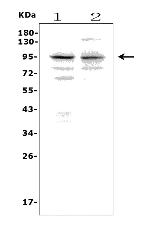
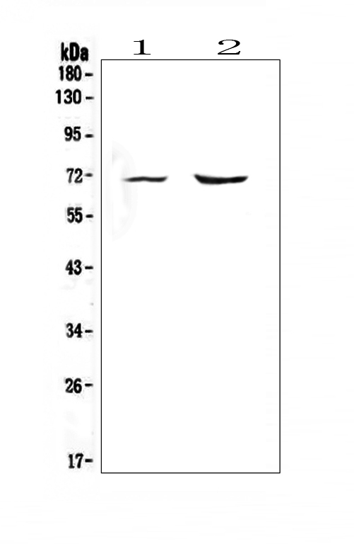
Figure 1. Western blot analysis of Pmel using anti-Pmel antibody (A01262-2).
Electrophoresis was performed on a 5-20% SDS-PAGE gel at 70V (Stacking gel) / 90V (Resolving gel) for 2-3 hours. The sample well of each lane was loaded with 50ug of sample under reducing conditions.
Lane 1: rat C6 whole cell lysates,
Lane 2: mouse brain tissue lysates.
After Electrophoresis, proteins were transferred to a Nitrocellulose membrane at 150mA for 50-90 minutes. Blocked the membrane with 5% Non-fat Milk/ TBS for 1.5 hour at RT. The membrane was incubated with rabbit anti-Pmel antigen affinity purified polyclonal antibody (Catalog # A01262-2) at 0.5 μg/mL overnight at 4°C, then washed with TBS-0.1%Tween 3 times with 5 minutes each and probed with a goat anti-rabbit IgG-HRP secondary antibody at a dilution of 1:5000 for 1.5 hour at RT. The signal is developed using an Enhanced Chemiluminescent detection (ECL) kit (Catalog # EK1002) with Tanon 5200 system. A specific band was detected for Pmel at approximately 95KD. The expected band size for Pmel is at 70KD.
Protein Target Info & Infographic
Gene/Protein Information For PMEL (Source: Uniprot.org, NCBI)
Gene Name
PMEL
Full Name
Melanocyte protein PMEL
Weight
70.255kDa
Superfamily
PMEL/NMB family
Alternative Names
D12S53EP1; gp100; ME20; ME20-M; melanocyte protein mel 17; Melanocyte protein Pmel 17; Melanocytes lineage-specific antigen GP100; Melanoma-associated ME20 antigen; melanosomal matrix protein17; PMEL17P100; premelanosome proteinME20M; SI; SIL; silver (mouse homolog) like; silver homolog (mouse); Silver locus protein homolog; silver, mouse, homolog of; SILVPmel17 PMEL D12S53E, ME20, ME20-M, ME20M, P1, P10017, SI, SIL, SILV, gp100, PMEL premelanosome protein melanocyte protein PMEL|melanocyte protein Pmel 17|melanocyte protein mel 17|melanocytes lineage-specific antigen GP100|melanoma-associated ME20 antigen|melanosomal matrix protein17|silver locus protein homolog|silver, mouse, homolog of
*If product is indicated to react with multiple species, protein info is based on the gene entry specified above in "Species".For more info on PMEL, check out the PMEL Infographic

We have 30,000+ of these available, one for each gene! Check them out.
In this infographic, you will see the following information for PMEL: database IDs, superfamily, protein function, synonyms, molecular weight, chromosomal locations, tissues of expression, subcellular locations, post-translational modifications, and related diseases, research areas & pathways. If you want to see more information included, or would like to contribute to it and be acknowledged, please contact [email protected].
Specific Publications For Anti-Melanoma gp100/Pmel Picoband™ Antibody (A01262-2)
Hello CJ!
No publications found for A01262-2
*Do you have publications using this product? Share with us and receive a reward. Ask us for more details.
Recommended Resources
Here are featured tools and databases that you might find useful.
- Boster's Pathways Library
- Protein Databases
- Bioscience Research Protocol Resources
- Data Processing & Analysis Software
- Photo Editing Software
- Scientific Literature Resources
- Research Paper Management Tools
- Molecular Biology Software
- Primer Design Tools
- Bioinformatics Tools
- Phylogenetic Tree Analysis
Customer Reviews
Have you used Anti-Melanoma gp100/Pmel Picoband™ Antibody?
Submit a review and receive an Amazon gift card.
- $30 for a review with an image
Be the first to review Anti-Melanoma gp100/Pmel Picoband™ Antibody
*The first user to submit a review for a product is eligible for Boster's Innovating Scientists Reward, which gives product credits. This is in addition to the gift card reward.
Customer Q&As
Have a question?
Find answers in Q&As, reviews.
Can't find your answer?
Submit your question
